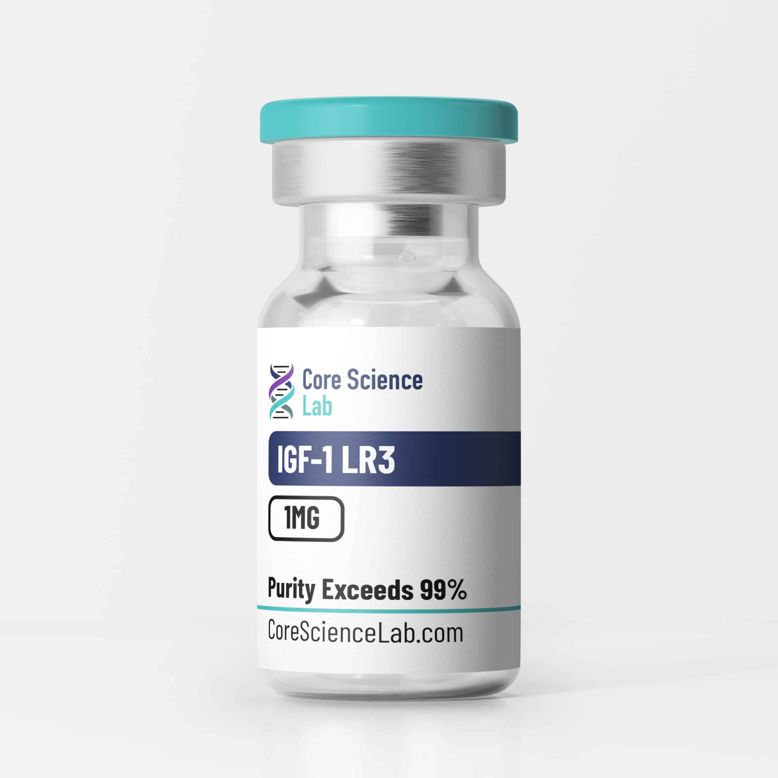
A high-purity, lyophilized synthetic analogue of human Insulin-like Growth Factor-1 (IGF-1). Characterized by an 83-amino acid sequence designed to evade Insulin-like Growth Factor Binding Proteins (IGFBPs), it is extensively utilized for research applications involving cellular proliferation, myogenesis, and targeted activation of the IGF-1 receptor (IGF-1R) signaling cascade.
Trust & Quality Verification
Purity Standard
Verified Analysis
Available
Shipping
IGF-1 LR3 (Long Arg3 Insulin-like Growth Factor-1) is a synthetic, engineered analogue of endogenous human IGF-1. Native IGF-1 is a 70-amino acid peptide that acts as the primary mediator of the somatotropic (growth hormone) axis. However, in physiological and in vitro settings, the bioavailability and half-life of native IGF-1 are heavily restricted by a family of six carrier proteins known as IGFBPs.
To overcome this regulatory bottleneck in experimental models, IGF-1 LR3 was synthesized with specific structural modifications: the addition of a 13-amino-acid extension at the N-terminus and the substitution of an arginine (Arg) for a glutamic acid (Glu) at position 3. These alterations drastically reduce the peptide’s binding affinity for secreted IGFBPs while preserving its high binding affinity for the IGF-1 receptor (IGF-1R). Consequently, IGF-1 LR3 exhibits significantly extended metabolic stability and up to a three-fold increase in biological potency compared to native IGF-1, making it a highly preferred reagent in mammalian cell culture, stem cell maintenance, and tissue regeneration assays.
Biochemical Characteristics
Chemically, IGF-1 LR3 functions as an elongated polypeptide engineered for uninhibited receptor interaction.
Chemical Properties
| Property | Specification |
| Molecule Name | IGF-1 LR3 |
| Synonyms | Long Arg3 IGF-1; Long R3 IGF-I |
| Molecular Weight | ~9111 g/mol |
| Form | Lyophilized Powder |
| Purity | ≥99% (Verified via HPLC) |
| Solubility | Soluble in water and organic solvents (refer to SDS) |
| Documentation | COA available per lot; SDS available |
(Note: Exact Molecular Weights and Formulas should be verified per lot COA/SDS).
IGF-1 LR3 is strictly for laboratory research and is commonly employed in the following investigational areas:
Mammalian Cell Culture & Stem Cell Maintenance
IGF-1 LR3 is extensively utilized as a highly potent supplement in serum-free media formulations for pluripotent stem cells, mesenchymal stem cells, and recombinant protein-producing cell lines (such as CHO cells). Researchers employ this peptide to maximize cellular survival, maintain undifferentiated states, and enhance cell density without the interference of endogenous binding proteins secreted by the cultured cells.
Myogenesis & Tissue Regeneration Models
In experimental models of tissue injury or mechanical overload, IGF-1 LR3 is used to study the autocrine and paracrine pathways governing cellular repair. Assays focus on quantifying the dose-dependent stimulation of skeletal myoblast proliferation, satellite cell activation, and the preservation of lean tissue mass independent of the systemic growth hormone axis.
Metabolic & Somatotropic Assays
Because it bypasses IGFBP regulation, IGF-1 LR3 serves as a precise chemical probe to isolate the direct effects of IGF-1R activation on cellular metabolism. Researchers investigate its impact on glucose uptake, amino acid transport, and the downstream modulation of endogenous growth hormone and insulin secretion.
Pathway / Mechanistic Context
The primary mechanism of action for IGF-1 LR3 in research settings revolves around the uninhibited, targeted activation of the IGF-1 receptor.
Preclinical Research Summary
Published preclinical literature documents investigations of IGF-1 LR3 across multiple experimental models:
Form & Analytical Testing
This material is produced via robust chemical synthesis and supplied as a lyophilized (freeze-dried) powder.
Referenced Citations
References are provided for informational purposes only and are not clinical claims.
RESEARCH USE ONLY
This product is intended strictly for laboratory research use only. It is not for human or veterinary use. It is not intended for diagnosis, treatment, cure, or prevention of any disease. All purchases are subject to our Terms of Service and Purity Guarantee.
No COAs available for this product.
RESEARCH USE ONLY
This product is intended strictly for laboratory research use only. It is not for human or veterinary use. It is not intended for diagnosis, treatment, cure, or prevention of any disease. All purchases are subject to our Terms of Service and Purity Guarantee.
Reviews
There are no reviews yet.